

Diagnostic Auxiliary Services
The Lomas Hospital, aware of the importance of having the best diagnostic support, puts at the disposal of the Medical Community and its patients, the most advanced equipment in the Region as well as a prestigious group of specialists in our Diagnostic Auxiliary Services.
RADIOLOGY AND IMAGE UNIT

The Radiology and Image Unit of the Lomas Hospital has been conceived since its inception as an area that, from its architecture, meets all the guidelines of international standards for areas of its type. Its equipment is a cutting edge technology as well as its organization, being a fully digitized department and that also has long term image storage systems in electronic format. The above allows the review of studies and sending them electronically to the different areas of hospitalization, outpatient consultation or outside the hospital via Internet, in order to meet all the expectations of the medical community and population in general.
The Radiology and Image Unit is constituted by the following services:
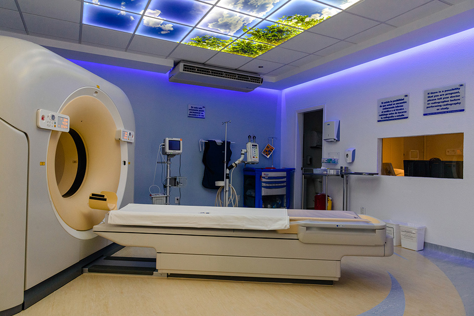
a) Multislice Tomography (128 cuts).
It offers high resolution benefits, studies with low radiation dose, faster and better resolution of image, taking care of your health in the warmest atmosphere.
Benefits:
• Reduce up to 60% of radiation.
• Greater study speed.
• Lower contrast volume.
• It has intelligent protocols that reduce radiation in sensitive areas such as:
o Eyes
o Thyroid
o Breast
o Lungs
o Genitals
• Special pediatric protocols with minimal radiation.
• Important increase in the resolution of images.
• Greater speed and accuracy in the post-processing of images.
• Advanced protocols for studies of:
o Heart
o Brain
o Body arteries
o Colon
b) Hemodynamics Unit
Unique equipment in the center of the country with digital flat detector that allows the obtaining of digital images of invasive heart studies with a resolution up to 30% more than conventional equipment of its type and that in addition to having a variable geometry, has applications for vascular studies of other parts of the body such as neurological applications, thorax, abdominals, extremities, etc..
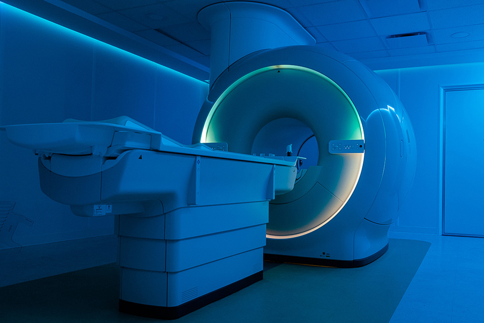
c) Magnetic Resonance
High magnetic field equipment that in addition to studies with application in neurology, thorax, abdomen and skeletal muscle system, has the most current technology such as spectrography and tractography.
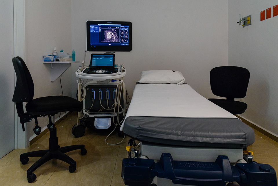
d) Echocardiography
The echocardiography team has the most current technology for the ultrasonographic study of the heart in two-dimensional form, with Doppler echo, in addition to being equipped with a trans esophageal transducer that allows a more precise intra-cavitary visualization of the heart and with the dobutamine module for cardiac stress tests with echocardiography in order to comprehensively assess the heart.
e) Digital mammography with tomosynthesis.
We have an equipment capable of performing high definition digital mammography and breast tomosynthesis; which consists of the acquisition of multiple images of low radiation in 3D for a deeper analysis of the mammary tissue.
Mammary gland biopsy system
Computer guided stereotactic biopsy system in both 2Dn and tomosynthesis systems.
Biopsies with margin of error less than 1 mm.
In addition, vacuum biopsy system and more samples with:
• Greater comfort
• Minimal invasion
• Greater speed
With probability of doing ambulatory or short stay procedures.
High definition ultrasound with Elastography Analysis
Specific for the study of small parts such as: thyroid, testicles, skeletal muscle and especially mammary glands.
It has capacity for elastography.
• Linear transducer for vascular study.
• Small head transducer for pediatric and neonatal patients.
• Multifrequency sector transducer with liver elastography capacity.
• Endocavitary multipurpose transducer.
New generation of matrix transducers for 3D and 4D multiplanar abdominal and obtetric studies.
An additional detection tool
A new technology called breast tomosynthesis has been developed and it has shown through clinical studies to be superior to conventional digital mammography.
What happens during the exam
A tomosynthesis test is performed as a traditional mammogram having many more benefits. The radiologist will guide you how to adjust, compress the breast under a pallet and take images from different angles. A tomosynthesis test is another tool in the early detection of breast cancer.
The study time is practically the same as in a conventional digital study and the obtaining of the images is almost immediate, which allows to assess at the moment and determine the need or not to make special shots.
“All women should have a breast cancer screening test once a year.”
f) Radiology and Fluoroscopy DuoDiagnost
There are two radiology teams in the Unit, for conventional and fluoroscopic studies with cutting edge equipment that allow the digitalization of images. There is also a conventional radiology room installed in the Emergency Unit in order to provide emergency care to patients who require it. There are two mobile x-ray equipment for use in different areas of hospitalization and a mobile arch for transoperatory fluoroscopic studies, with storage and sending of digital images.
g) Ultrasound 4D
It has Doppler Color Ultrasound equipment with convex and linear multifrequency transducers, as well as endocavitary transducer, which allow to perform all kinds of studies with high resolution images, as well as abdominal, pelvic, transfontanellar, of small parts, vascular, and pregnancy studies with technology in third and fourth dimensions covering a wide range of clinical applications, including biopsies, intra-hospital and trans operatory portable studies.
CLINICAL DIAGNOSIS AND CHECK UP UNIT
Unit where auxiliary diagnostic studies are carried out in ambulatory patients, such as cardiopulmonary evaluation and lung function tests, spirometry tests, electrocardiography ambulatory register (holter) and ambulatory blood pressure are performed.
Its objective is to carry out preventive check up studies that include clinical evaluation of cardiovascular and respiratory risk, general laboratory studies, imaging studies: chest x-ray, mammography, ultrasound, densitometry and audiometry, according to the executive check up packages that we have.

CLINICAL LABORATORY AND BLOOD BANK
For the clinical analysis, we have a Clinical Laboratory with almost 6 decades of experience, equipped with the latest generation equipment and a cutting edge technology, which provides service 24 hours a day, 365 days of the year.
The laboratory has distinguished itself for its technological vanguard and excellence in service, which has allowed it to obtain national recognition by offering its patients and maquila services, timely services and reliable results in a consistent manner.
We cover all areas of the clinical laboratory such as Hematology, Chemistry, Infectology, Endocrinology and Immunology as well as special tests.
In the area of microbiology we have the safest and most reliable diagnostic platform because it uses a MALDI TOF device, a team that uses mass spectrophotometry to analyze at the molecular level the identification of micro organisms.
We are associated with the most prestigious international laboratories so that we can attend all specialized analysis requests that are not made locally.
The Blood Bank, attached to the Clinical Laboratory, has all the latest generation diagnostic tests and also works 365 days a year for each of the blood donors and recipients to obtain a safe blood donation, In addition to offering special procedures such as Plaquetpheresis and Therapeutic Bleeding.

PATHOLOGICAL ANATOMY DEPARTMENT
The Department of Pathological Anatomy of Lomas de San Luis Hospital is a department with more than 30 years of experience, with highly qualified personnel and at the forefront of histopathological diagnostic studies. It is recognized in the field of pathology for its career and performance; for example its active academic participation that has been something that has distinguished it in its trajectory. It has the interrelation with other departments of Pathology, both national and international, that allows a greater diagnostic precision in its interaction.
The Laboratory has two medical specialists in pathological anatomy, a cytotechnologist, a Histotechnologist, a Molecular Biologist, a Secretary, an Administrator and a Director of Quality and Continuous Improvement.
Among the services offered are the following:
SURGICAL PATHOLOGY.
“The first step and the basis for a specific treatment, is a precise diagnosis”.
The pathological study of biopsies and surgical pieces is fundamental to determine the diagnosis of multiple diseases, especially cancer. Our Hospital has a Department of Pathological Anatomy with microscopy equipment, macroscopy unit, tissue processing, microtomy, photomicroscopy, as well as the use of importing reagents of the highest quality. All surgical pieces with cancer are staged according to the most recent protocols of the College of American Pathologists and the American Joint Committee on Cancer. We have a network of world class experts from the best hospitals and universities in the USA as consultants in cases of extreme diagnostic difficulty.
1. IMMUNOHISTOCHEMISTRY.
Undoubtedly, the technique that has revolutionized the pathological diagnosis in the last 50 years is Immunohistochemistry. This powerful tool is absolutely indispensable at the present time in the pathology laboratories, since it allows to corroborate, establish or refine multiple diagnoses, especially cancer cases, as well as to determine the existence or not, of prognostic and predictive factors of response to treatments “white€. Our laboratory has more than 160 antibodies (and is constantly expanding), uses the most modern systems worldwide and a 27 year experience -as well as specific training- by the Department Director in this technique.
2. CITOPATHOLOGY.
Our laboratory has a gynecological cytology interpreting service (Papanicolaou) with a liquid based system, which is the method used in the US and Europe for the timely detection of cervico-uterine cancer. Also of cytologies of organic liquids (expectoration, pleural fluid, peritoneal, urine, etc.) and specimens of Puncture Aspiration with Fine Needle (thyroid gland, salivary glands, mammary gland, lymphatic nodes,etc.).
3. MOLECULAR BIOLOGY.
The laboratory offers molecular biology services, necessary for the prediction of response to treatments against various types of cancer, as well as for diagnostic purposes. To do this, we have a collaboration agreement with a reference laboratory with the highest standards in this technique, as long as we are assembling it in our laboratory.